Next to periodontitis, caries are considered the most common dental disease. They are caused by a combination of bacteria and sugar. Depending on the type and progress of the disease, caries can be very painful for patients. It is therefore important to detect and treat caries at an early stage. This article will look into various options for caries detection.
Development of caries
Each type of caries is caused by bacteria in dental plaque. Bacteria metabolize sugars from consumed food and produce acid that can attack and damage the enamel. Generally, the earlier this is treated, the less tooth structure is lost.
The consequences, detection, and treatment of caries strongly depend on the type of bacteria, oral hygiene, dietary habits, and also the degree of mineralization/hardness of the teeth. There are types of caries that are largely inactive for a long time and types that progress very quickly and aggressively. Depending on the type, there are different methods for detecting carious lesions.
Clinical examination
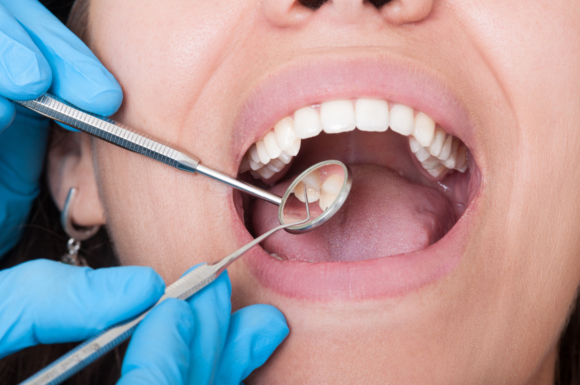
During the annual routine check-up at the dentist, the teeth are always examined for caries. This visual or clinical examination is performed using good lighting and a special mirror. During this procedure, the dentist examines superficial discolorations and carious lesions in order to make an initial diagnosis. However, this method does not provide an adequate examination of interdental spaces.
Radiological diagnostics
X-ray is probably the most common method for caries diagnosis. Bitewing radiographs or panoramic radiographs can be used to detect enamel lesions in particular. Thus, the carious lesions in the interdental spaces can be detected very reliably (up to 90 percent). During radiological diagnostics, patients are exposed to very small amounts of X-radiation. Therefore, an X-ray is always based on an examination and indication that justify this treatment.
3D X-ray imaging with CBCT
One development in radiological diagnostics is 3D X-ray. Cone beam computed tomography (CBCT) provides more reliable results than conventional 2D X-ray. However, 3D X-ray is generally used only when the results of 2D X-ray are not sufficient. This is mainly due to the higher costs of CBCT, which patients usually have to bear themselves.
Laser-based caries diagnostics
Laser-based caries diagnostics is also known as laser fluorescence caries detection. Using a special laser fluorescence device, light is absorbed by organic and inorganic substances. Areas affected by caries are stimulated to fluoresce, which ultimately results in an acoustic signal. Thus, the dentist can determine whether or not there is a carious lesion. This method is particularly suitable for detecting occlusal caries.
Caries detector
The caries detector is usually only used in the context of a pending excavation, i.e. the removal of carious dentin. Caries are made visible by a chemical dye. The closer it gets to the pulp, the darker the color. However, this also increases the risk of opening the pulp chamber, which can be painful for patients.
Fiber-optic transillumination
Similar to laser-based caries diagnostics, fiber-optic transillumination also uses a powerful light source. In this case, cool white light transmits the tooth. Healthy and carious structures show different light refraction behavior. This makes it easy to distinguish between the two states. Carious areas are perceived as dark shadows. This approach is particularly useful for detecting dentin caries.
AI-powered software
The latest developments and findings are emerging in the field of artificial intelligence. Using software from dentalXrai, X-ray images are evaluated by a specially developed AI which detects caries within seconds. Among other things, carious lesions are color-coded so that dentists can quickly and reliably analyze the results and use them for subsequent patient communication. The software thus serves as a supporting tool in radiological diagnostics. AI can assist especially in the area of early detection. According to the results of the 2020 study “Detecting caries lesions of different radiographic extension on bitewings using deep learning”, AI detects up to three times more early carious lesions compared to conventional diagnostics conducted by dentists.
