Fully automated X-ray diagnosis
with artificial intelligence (AI) developed at Charité Berlin
- Better communication with patients
- Highest medical quality
- Create written documentation automatically
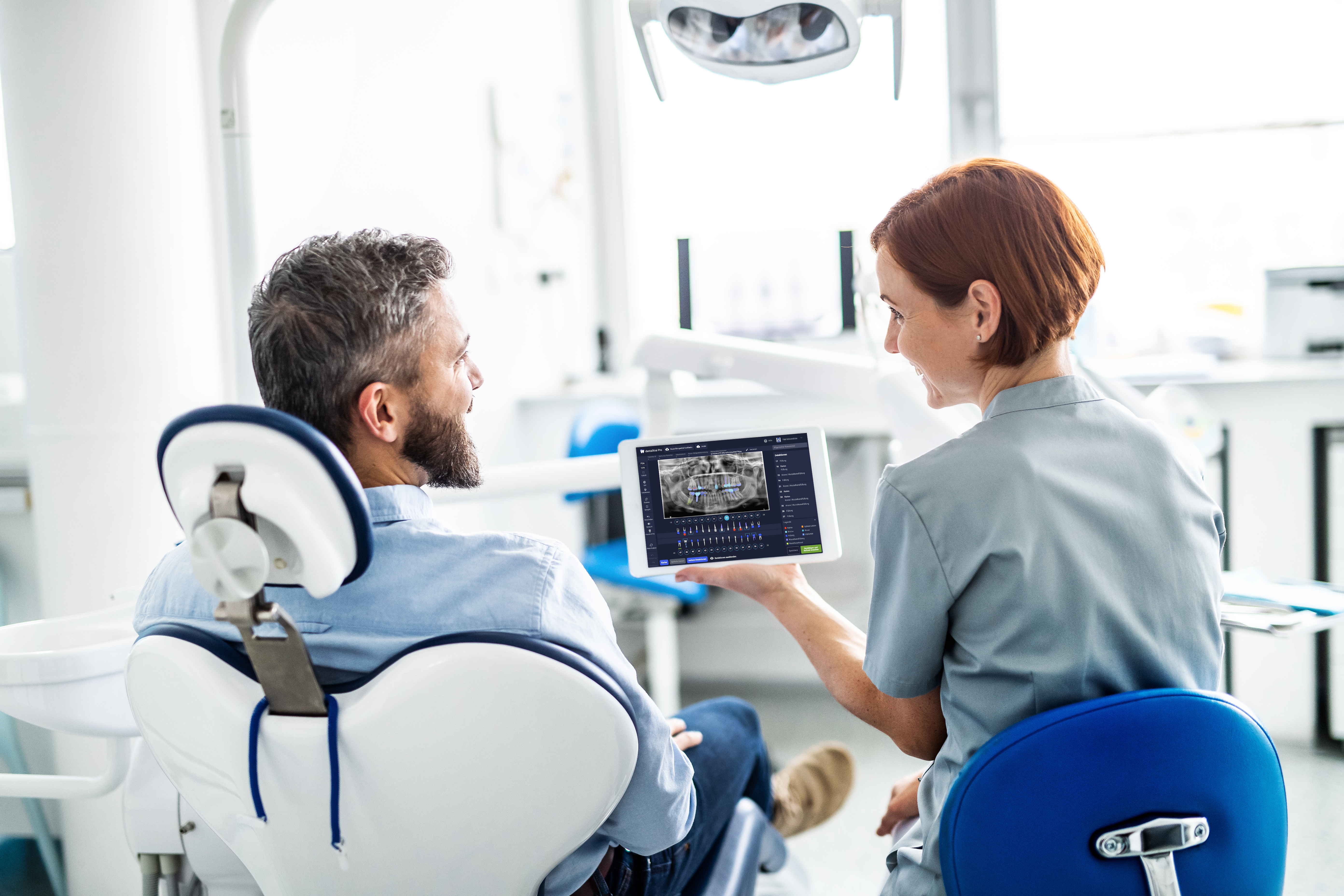
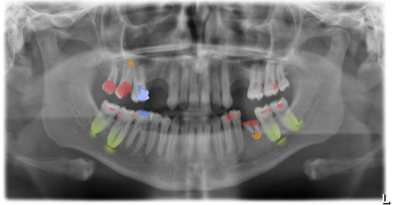
The software detects caries and infections, but also implants and root fillings with reliably high quality. The findings are highlighted in color for better visualization and dentalXrai automatically generates written documentation, saving dentists valuable time and improving patient communication.
dentalXrai with infoskop® in use at ZahnOase in Berlin

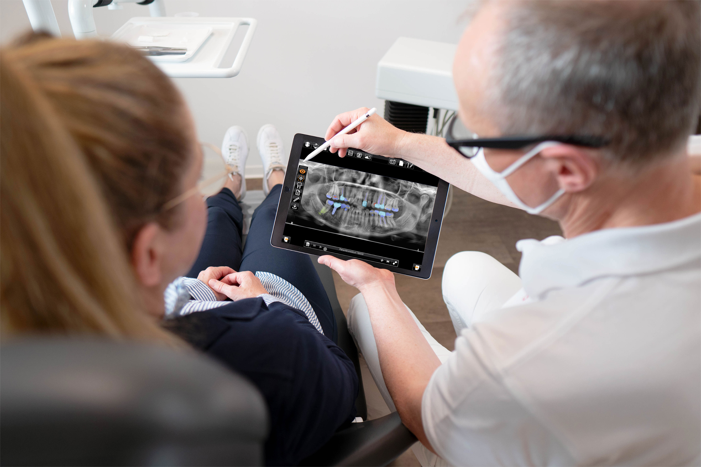
Figure: dentalXrai as part of the infoskop® software on the iPad
Using dentalXrai means:
- Detect lesions at an early stage
- Reduce the time required
- Create findings report automatically
- Optimize therapy recommendations
dentalXrai in everyday practice
Diagnostics and written findings at the “push of a button”
The integration into the existing everyday practice infrastructure is also ideally implemented as a merger with existing practice management systems. Independent of the manufacturer, we offer different integration models so that dentalXrai could be implemented with all practice software and X-ray equipment.
- Fully automated data exchange with AI
- Less misdiagnosis and better patient communication
- More confidence in your therapy recommendation
- Perfect documentation
Three steps to the diagnosis with dentalXrai
1. Acquire X-ray image and transfer automatically to dentalXrai
2. Obtain dentalXrai diagnostics, adjust detection if necessary and confirm analysis.
3. Discuss diagnosis and therapy recommendation with patient

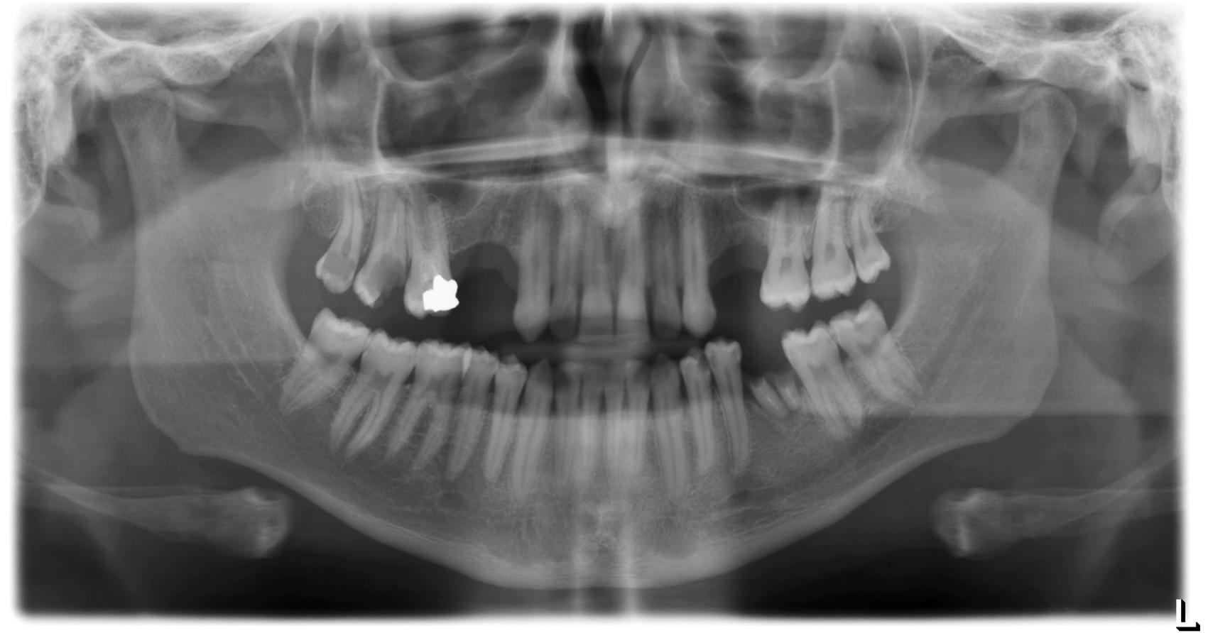
Figure: native X-ray image without detections

Figure: X-ray image with detections by dentalXrai
The integrated control bar allows the operator to switch back and forth between the native X-ray image and the color-highlighted detections. This also makes it easy for the patient to understand the diagnosis and treatment plan.
Systematic and detailed documentation enables progress monitoring, supports therapy planning and also forensically safeguards the findings. The written documentation is stored in the everyday practice software.

Figure: Schematic representation with exemplary findings of dentalXrai
We look forward to introducing dentalXrai to you!
Learn about our AI solution for dental professionals
- Automated processes
- Time-saving support
- Color coded detections

dentalXrai: AI for dental practices
- Trained for panoramic slice exposures and bite wing exposures
- Time-saving AI support in daily practice routine
- Developed for the needs of dentists
Learn more about dentalXrai’s AI technology here:
Our partners


