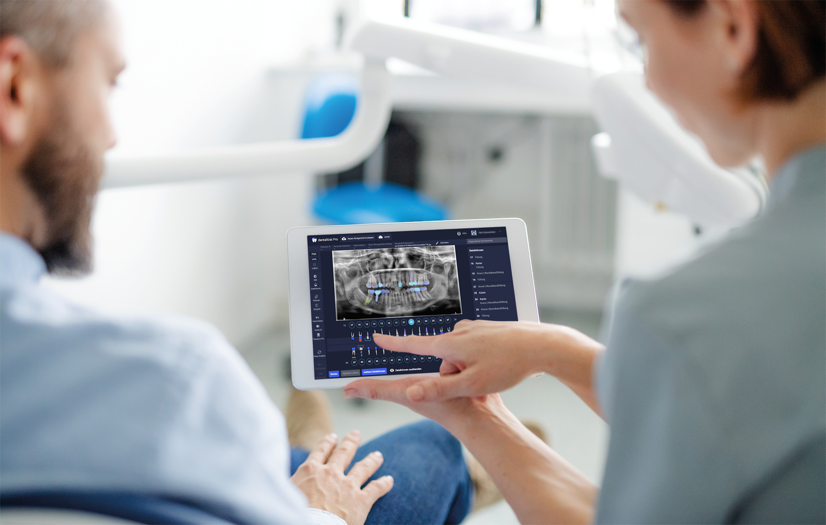
Artificial intelligence (AI) has come a long way in recent years. There are many areas of application and AI has now also arrived in dentistry. To support dentists in their daily work, to save time, and simultaneously improve patient communication, dentalXrai has developed an AI-based software.
What is dentalXrai?
Many patients have certainly already had an X-ray taken at the dentist’s at least once. During the subsequent consultation and explanation of the findings, comprehension problems frequently arise. In most cases, patients will not understand their X-ray images. In the end, the consultation is a matter of trust—patients have to rely on the dentist’s explanations.
DentalXrai solves this problem, as well as many others. The idea for the start-up was born in 2017 under the leadership of Prof. Dr. Falk Schwendicke and Dr. Joachim Krois. It was spun off from Charité Berlin in April 2020. The AI-based software recognizes and classifies teeth and detects pathologies (e.g. caries, apical lesions) as well as non-pathological structures (restorations). To achieve this, the dentalXrai team trained artificial neural networks with tens of thousands of annotated X-ray images. The AI has been trained especially on panoramic and bitewing radiographs. Through continuous improvement and updates of the models, it has been proven to achieve better and more reliable results than those of many dentists.*
An existing 2D X-ray image of the intraoral space (mouth) is uploaded to the software either automatically or manually. In just a few seconds, dentalXrai automatically evaluates the image. The dentist receives the results and can adjust the detections if necessary. Detections are highlighted in color, which significantly improves the already mentioned communication with the patient. Through AI, patients can now visually understand their dentists’ findings. In addition, the results are digitally processed so that it is possible to switch between the color-coded and original image.
The advantages of X-ray analysis with AI
Probably the most important advantages for dentists are time savings and improved patient communication. Fully automated AI X-ray reporting within seconds leaves more time for discussing findings with your patient. In addition, the results are automatically transferred to a digital tooth chart. This means less documentation effort and optimal integration into digital dental practices. Findings can also be explained and understood much more easily and comprehensibly.
The software dentalXrai Pro supports dentists with an additional expert opinion in their daily work. This also means that there are fewer false diagnoses. The AI-based findings are more reliable than conventional analyses of X-ray images with the naked eye.*
The advantages of dentalXrai at a glance:
- evaluation of dental X-ray images within seconds
- better patient communication through color-coding
- fewer false diagnoses
- automatic report on findings
- trained for panoramic and bitewing radiographs
- great support for daily work
- instant digital documentation
* According to the study “Detecting caries lesions of different radiographic extension on bitewings using deep learning” of 2020 our AI – dentalXrai Pro – detects up to 3x more caries lesions at an early stage in comparison with conventional dental diagnoses.
