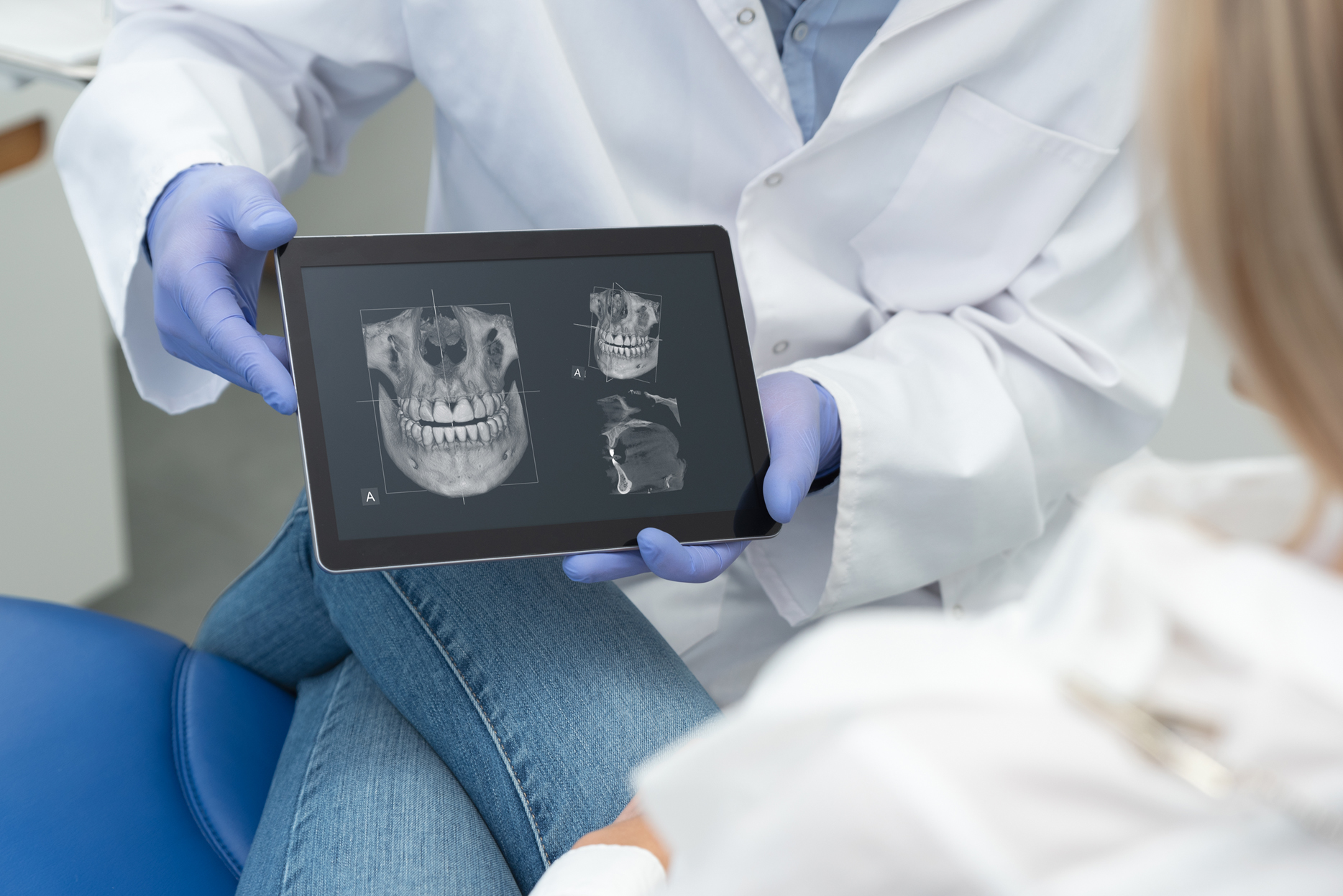
The use of X-ray imaging is commonplace in dental practices. For decades, X-ray images have been a great support for dentists to detect as well as treat diseases in the maxillofacial region. 3D X-ray imaging has become more and more widespread for several years. Here you will find out what it is all about: how does it differ from conventional X-ray imaging and when does it make sense to use 3D cone beam computed tomography (CBCT)?
What is 3D X-ray imaging?
X-rays are indispensable for dentists in their daily work. Only about one third of the tooth and the surrounding structure are visible to the human eye. Accordingly, X-rays are an essential tool for diagnosis. For example, tooth anatomy, position, condition of the nerves, the length of the roots, or even chronic inflammations are only visible in this way.
X-rays are often associated with X-radiation. However, radiation levels are significantly lower than patients assume. The natural radiation surrounding us every day is about 10 µSv (microsievert). The radiation of one X-ray exposure, on the other hand, is about 5 µSv.
X-ray imaging has already been used for decades. In recent years, 3D X-rays have also become increasingly popular among dentists. This is a further development of 2D X-ray imaging and offers faster as well as more precise X-rays. The dentist can thus view the jaw from different angles. The X-ray scanner rotates around the patient’s head and takes hundreds of images within a few seconds. At the end of the scanning process, these are assembled to form a holistic three-dimensional image.
Advantages and disadvantages compared to 2D X-ray imaging
First of all, a distinction should be made between the conventional X-ray procedure and digital 2D X-ray imaging. With the latter, radiation exposure is up to 80 percent lower. Instant digitization makes it easier to discuss the results with patients. Here, it is also advantageous that the image quality is significantly higher. AI-powered software such as dentalXrai can also process and analyze digital X-ray images. The images can even be evaluated directly on the iPad®.
Nevertheless, many findings that would remain undetected by the 2D procedure can be identified by cone beam computed tomography (CBCT). With 3D technology, the patient’s anatomical features are displayed spatially. It therefore also offers greater planning reliability for surgical interventions. With 3D X-ray imaging, the diagnosis is also easier for patients to understand due to spatial visualization. Last but not least, CBCT results in lower radiation exposure.
Only one question remains: why aren’t all practices upgrading to 3D X-ray imaging? 3D X-rays are simply not necessary for every patient. They are rather a supplement and are often only used when conventional x-rays are inadequate or do not provide the desired results. A CBCT machine is also very expensive to purchase; a fact patients will notice. The costs for CBCT images are usually not covered by health insurance and can range from €100 to €300.
Conclusion: when do dentists use 3D X-ray imaging?
CBCT is never a replacement for conventional or digital 2D X-ray imaging. It rather plays a supporting role at the dentist’s. Therefore, if 2D X-ray images are not sufficient for reliable diagnosis or planning of treatment, it is possible to resort to the more modern option. However, costs are an important point. For the dental practice, CBCT is very expensive to purchase. The patients also have to pay extra, since CBCT scans are not usually covered by health insurance.
